Jorge Cardoso wears many hats, and that’s appropriate given he has so many brains. A hundred thousand of them to be exact.
Cardoso is a teacher, a CTO, an entrepreneur, a founding member of the MONAI open source consortium and a researcher in AI for medical imaging. In that last role, Cardoso and his team just discovered ways to create realistic, high resolution 3D images of human brains with AI.
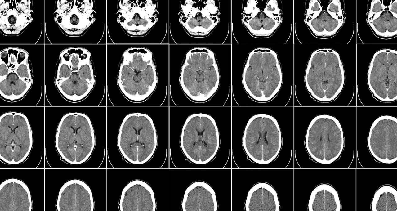
The researcher at King’s College London and CTO at the London AI Centre is making 100,000 synthetic brain images available free to healthcare researchers. It’s a treasure trove that could accelerate understanding of dementia, aging or any sort of brain disease.
Accelerating AI in Healthcare
“In the past, many researchers avoided working in healthcare because they couldn’t get enough good data, but now they can,” said Cardoso.
“We want to direct the energy of AI research into healthcare,” he said.
It’s a major donation compared to the world’s largest repository of freely available brain images. The UK Biobank currently maintains multiple brain images taken from more than 50,000 participants, curated at an estimated cost of $150 million.
Synthetic Data for Science
The images represent an emerging branch in healthcare of synthetic data, something that’s already widely used in computer vision for consumer and business apps. Ironically, those fields also have access to open datasets with millions of real-world images.
By contrast, medical images are relatively scarce, typically only available to researchers connected to large hospitals, given the need to protect patient privacy. Even then, medical images tend to reflect the demographics the hospital serves, not necessarily the broader population.
A fortunate feature of the new AI approach is it can make images to order. Female brains, male brains, old ones, young ones, brains with or without disease. Plug in what you need, and it creates them.
Though they’re simulated, the images are highly useful because they preserve key biological characteristics, so they look and act like real brains would.
Scaling with MONAI on Cambridge-1
The work required a supercomputer running super software.
NVIDIA Cambridge-1, a supercomputer dedicated to breakthrough AI research in healthcare, was the engine. MONAI, an AI framework for medical imaging, provided the software fuel.
Together they created an AI factory for synthetic data that let researchers run hundreds of experiments, choose the best AI models and run inference to generate images.
“We couldn’t have done this work without Cambridge-1 and MONAI, it just wouldn’t have happened,” Cardoso said.
Massive Images, Up to 10x Speedups
An NVIDIA DGX SuperPOD, Cambridge-1 packs 640 NVIDIA A100 Tensor Core GPUs, each with enough memory to process one or two of the team’s massive images made up of 16 million 3D pixels.
MONAI’s building blocks include domain-specific data loaders, metrics, GPU accelerated transforms and an optimized workflow engine. The software’s smart caching and multi-node scaling can accelerate jobs up to 10x, said Cardoso.
He also credited cuDNN and “the whole NVIDIA AI software stack that helped us work much faster.”
Beyond the Brain
Cardoso is working with Health Data Research UK, a national repository, to host the 100,000 brain images. The AI models will be available, too, so researchers can create whatever images they need.
There’s more. The team is exploring how the models can make 3D images of any part of the human anatomy in any mode of medical imaging — MRIs, CAT or PET scans, you name it.
“In fact, this technique can be applied to any volumetric image,” he said, noting users may need to optimize the models for different types of images.
Many Directions Ahead
The work points to many directions Cardoso described enthusiastically as if unloading the contents of multiple minds.
Synthetic images will help researchers see how diseases evolve over time. Meanwhile his team is still exploring how to apply the work to body parts beyond the brain and what kinds of synthetic images (MRI, CAT, PET) are most useful.
The possibilities are exciting and, like his many roles, “it can be a bit overwhelming,” he said. “There are so many different things we can start thinking about now.”