NVIDIA and Partners Build in America, for America
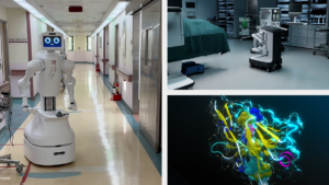
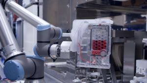
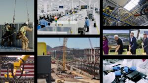
NVIDIA and its partners are investing in American manufacturing, supply chains, energy grids and skilled workforces so the U.S. can produce the infrastructure needed for better healthcare, breakthrough scientific discovery,...