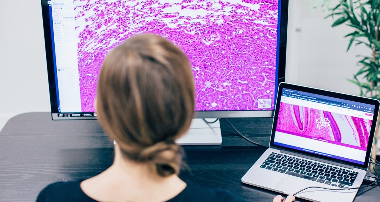
Pathology, the study and diagnosis of disease, is a growth industry.
As the global population ages and diseases such as cancer become more prevalent, demand for keen-eyed pathologists who can analyze medical images is on the rise. In the U.K. alone, about 300,000 tests are carried out daily by pathologists.
But there’s an acute personnel shortage, globally. In the U.S., there are only 5.7 pathologists for every 100,000 people. By 2030, this number is expected to drop to 3.7. In the U.K., a survey by the Royal College of Pathology showed that only 3 percent of histopathology departments had enough staff to meet demand. In some parts of Africa, there is only one pathologist for every 1.5 million people.
While pathologists are under increasing pressure to analyze as many samples as possible, patients are having to endure lengthy wait times to get their results.
Aiforia, a member of the NVIDIA Inception startup accelerator program, has created a set of AI-based tools to speed and improve pathology workflows — and a lot more.
The company, which has offices in Helsinki and Cambridge, Mass., enables tedious tasks to be automated and for complex challenges to be solved by unveiling quantitative data from tissue samples.
This helps pathologists overcome common obstacles to the development of versatile, scalable processes for medical research, drug development and diagnostics.
“Today we can already support pathologists with AI-assisted analysis, but AI can do so much more than that,” said Kaisa Helminen, CEO of Aiforia. “With deep learning AI, we are able to extract more information from a patient tissue sample than what’s been previously possible due to limitations of the human eye.
“This way, we are able to promote new discoveries from morphological patterns and facilitate more accurate and more personalized treatment for the patient,” she said.
Hidden Figures
AI has made it possible to automate medical imaging tasks that have traditionally proved nearly impossible for the human eye to handle. And it can reveal information that was previously hidden in image data.
With Aiforia’s AI tool assisting the diagnostic process, pathologists can improve the efficiency, accuracy and reproducibility of their results.
Its cloud-based deep learning image analysis platform, Aiforia Create, allows for the rapid development of AI-powered image analysis algorithms, initially optimized for digital pathology applications.
Aiforia initially developed its platform with a focus on cancer, as well as neurological, infectious and lifestyle diseases, but is now expanding it to other medical imaging domains.
For those who want to develop algorithms for a specific task, Aiforia Create provides domain experts with unique self-service AI development tools.
Aiforia trains its image analysis AI models using convolutional neural networks on NVIDIA GPUs. These networks are able to learn, detect and quantify specific features of interest in medical images generated by microscope scanners, X-rays, MRI or CT scans.
Its users can upload a handful of medical images at a time to the platform, which uses active learning techniques to increase the efficiency of annotating images for AI training.
Users don’t need to invest in local hardware or software — instead they can access the software services via an online platform, hosted on Microsoft Azure. The platform can be deployed instantly and scales up easily.
Unpacking Parkinson’s
Aiforia’s tools are also being used to improve the diagnosis of Parkinson’s disease, a debilitating neurological condition affecting around one in 500 people.
The disease is caused by a loss of nerve cells in a part of the brain called the substantia nigra. This loss causes a reduction in dopamine, a chemical that helps regulate the movement of the body.
Researchers are working to uncover what causes the loss of nerve cells in the first place. Doing this requires collecting unbiased estimates of brain cell (neuron) numbers, but this process is extremely labor-intensive, time-consuming and prone to human error.
University of Helsinki researchers collaborated with Aiforia to mitigate the challenges of traditional methods to count neurons. Brain histology images were uploaded to Aiforia Hub, then they deployed the Aiforia Create tool to quantify the number of dopamine neurons in the samples.
Introducing the computerized counting of neurons improves the reproducibility of results, reduces the impact of human error and makes analysis more efficient.
“It’s been studied so many times that if you send the same microscope slide to five different pathologists, you get different results,” Helminen said. “Using AI can help bring consistency and reproducibility, where AI is acting as a tireless assistant or like a ‘second opinion’ for the expert.”
The study carried out at the University of Helsinki would typically have taken weeks or even months without AI. Using Aiforia’s tools, the research team was able to achieve a 99 percent speed increase, freeing up their time to further their work into finding a cure for Parkinson’s.
Aiforia Create is sold with a research use status and is not intended for diagnostic procedures.
